Exploration of Antibacterial and Anti-inflammatory Activities of Premna integrifolia Plant Extracts in Bubaline Mastitis- Juniper Publishers
Juniper Publishers- Journal of Complementary Medicine
Abstract
Mastitis, which affects the milk production of dairy
animals, is usually due to mammary gland invasion by bacterial
pathogens. Emergence of antimicrobial resistance in bacteria and side
effects associated with the use of anti-inflammatory cortisones in
mastitis prompted for use of alternate/complementary therapeutics. As
the plant Premna integrifolia was reported to exhibit
antibacterial, anti-inflammatory/immunomodulatory properties, its leaf
and root aqueous extracts were tested for their antibacterial activity
against Staphylococcus aureus and Escherichia coli, either
individually or in combination with the antibiotics. The
anti-inflammatory properties of the extracts were also tested against
the bubaline mammary epithelial cells (MEC) infected with S. aureus and E. coli. In microbroth dilution assays for assessing minimum inhibitory concentration (MIC) in vitro, the leaf and root extracts of Premna integrifolia didn’t exhibit any antimicrobial activity against S. aureus but showed significant antimicrobial activity on E. coli. In combination with the plant extract, the sensitivity of S. aureus to amoxicillin is not only increased but also the S. aureus isolates that were resistant to amoxicillin also became sensitive. The Premna integrifolia
leaf and root extracts, however, showed antagonism on antimicrobial
activity of enrofloxacin in combination. In addition the aqueous root
extract of Premna integrifolia exhibited anti-inflammatory activity through down regulation of cytokines IL-6, IL-8 and TNF-α in S. aureus and IL-6 and IL-8 in E. coli infected MEC. These studies reveal antimicrobial activity of leaf and root extracts of Premna integrifolia on E. coli. In combination with amoxicillin these plant extracts increased the sensitivity of S. aureus to amoxicillin. The anti-inflammatory activity of root extract of Premna integrifolia on MEC infected with S. aureus and E. coli is also demonstrated in these studies.
Keywords:Mastitis; S. aureus; E. coli; Premna integrifolia; Mammary epithelial cells; cytokines; Amoxicillin; EnrofloxacinIntroduction
Mastitis in dairy animals is inflammatory reaction of
the udder tissue against the invading microbial pathogens. Bacterial
pathogens are majorly implicated in the mastitis of cows and buffaloes
leading to major production losses in dairy animals resulting in huge
economic losses to dairy farmers and industry [1]. Staphylococcus aureus and Escherichia coli
are the major bacterial pathogens of bovine/bubaline mastitis [2].
However, the emergence of antimicrobial resistance in bacterial
pathogens that cause mastitis in dairy animals is a cause of grave
concern [3-5]. Also controlling the inflammation in mastitis is very
essential as the persistent inflammation of mammary gland tissue may
result in permanent unproductivity in dairy animals [6-7]. Mastitis is
the most frequent reason for the use of antimicrobial drugs in dairy
herds, which eventually has resulted in antimicrobial resistance [8].
Development of new antibiotics will take long time
and there is chance of further developing antimicrobial resistance
against these molecules in due course. In this context exploration of
natural compounds from medicinal plants that exhibit both antibacterial
and anti-inflammatory/immunomodulatory properties may offer promising
solution for therapeutic approach to mastitis in dairy animals.
Medicinal uses of the plant Premna integrifolia that has
prominent value in Indian system of medicine Ayurveda was reviewed by
different researchers [9-11]. Reports on increased sensitivity of
bacterial pathogens to antibiotics, when used in combination with
anti-inflammatory compounds like Non-Steroidal Anti-inflammatory Drugs
(NSAIDs) [12], also encourages us to take up research work on the
natural compounds with anti-inflammatory activity. As the development of
resistance to natural products of plant origin is highly remote and the
issue of antibiotic residues in milk doesn’t arise with the natural
compounds, the present investigation was taken up to study the
antibacterial and anti-inflammatory/immunomodulatory activities of
aqueous young leaf and root extracts of the plant Premna integrifolia. The study is aimed to test the anti-bacterial activity of the plant extracts on S. aureus and E. coli, either individually or in combination with
the antibiotics. It is also aimed to test the anti-inflammatory/
immunomodulatory activity of the plant extracts on Mammary
epithelial cells (MEC) cultured from fresh milk of buffaloes and
further infected with the selected bacterial pathogens of mastitis.
Materials and Methods
Plant material
Plant materials were collected from Maharastra region of
India. The plant was identified as Premna integrifolia L. belonging
to Verbenaceae by Dr. S. K. Srivastava, Scientist-E, BSI, Dehradun
with accession no. 116123. Sample herbarium sheets deposited
with Northern Regional Centre, Botanical Survey of India,
Dehradun.
Preparation of Premna integrifolia extracts
The young roots and leaves of Premna integrifolia were sun
dried for 15 days, powdered and successively extracted with
soxhlet apparatus with petroleum ether, ethyl acetate, methanol
and water in the increasing polarity index. These extracts were
dried using a rotatory evaporator followed by lyophilization.
Similarly, leaves were dried in shade for 10 days and extracted as
above. In the present study the aqueous extracts were evaluated
for their anti-microbial and anti-inflammatory effects./p>
Bacterial isolates
The bacterial pathogens Staphylococcus aureus and Escherichia
coli were isolated from the mastitic milk samples of buffaloes
and the bacteria were subjected to characterization by culturing
on selective bacteriological media. Mannitol salt agar (MSA) and
Eosin methylene blue (EMB) agar (Oxoid, UK) were used for
culture of S. aureus and E. coli, respectively. These bacteria were
further characterized in polymerase chain reaction (PCR) test by
reactivity with species-specific oligonucleotide primers [2].
Microbroth dilution method for measuring the minimum inhibitory concentration (MIC) of antibiotic/ minimum inhibitory concentration (MIC) of antibiotic/
The antimicrobial activity of the plant extracts was evaluated
by microbroth dilution method in serial wells of microtitre plate
(Axygen, USA) [13], with suitable modifications. Briefly, two-fold
dilution of antibiotic/plant extract (10mg/ml) is made with cation
adjusted Mulleur Hinton broth, in their respective wells of 96-well
microtiter plate. The antimicrobial activity of the plant extracts
was tested individually, also in combination with antibiotic. In
the combination studies a fixed volume of 50μl of plant extract
(10mg/ml) was added to the wells with serial dilution of
respective antibiotic. Separate row(s) of wells with serial dilution
of antibiotic alone were also maintained to compare the MIC values
of antibiotic with the MIC values of plant extract or antibiotic
& plant extract combination. Appropriate controls were also
maintained. Amoxicillin and enrofloxacin (SRL, India) antibiotics
in powder form were used for S. aureus and E. coli, respectively. To
all the wells constant volume of 300μl of 0.5 McFarlands standard
bacterial culture (S. aureus/E. coli) was added. The culture plates
were incubated for 18hrs. and the absorbance readings were
taken at 660 nm (Multiskan plate reader, Thermo). The MIC values
of the antibiotic/plant extract or combination of antibiotic & plant
extract corresponding to the absorbance readings of respective
wells were noted. Then indicator dye p-iodonitrotetrazolium violet
(INT) (SRL, India) was added to all the wells to visually appreciate
the extent of antimicrobial activity of the compounds tested.
The breakpoints of amoxicillin and enrofloxacin/ciprofloxacin in
MIC assays were taken as per Clinical and Laboratory Standard
Institute (CLSI) guidelines 2012.
In Microbroth dilution method for measuring the MIC a loopful
of inoculum was picked up from the wells in microtiter plates
where there is inhibition of bacterial growth and streaked on
bacteriological medium, further incubated to confirm the absence
of live bacteria/bacterial growth in those wells.
Isolation and culture of mammary epithelial cells (MEC) from milch buffaloes
Mammary epithelial cells were isolated form the fresh milk
of apparently healthy milch buffaloes maintained at Livestock
Farm Complex, NTR College of Veterinary Science, Gannavaram
as per the established procedure [14] with suitable modifications.
Briefly, the fresh milk samples were centrifuged at 500 x g and the
cell pellet was washed with phosphate buffer saline (pH 7.2). Then
the cell pellet was cultured in DMEM/F12 (Sigma, USA) medium
with 10% Foetal Bovine Serum (Thermo Fisher) for 48 hrs. in 5%
CO2 atmosphere. Four groups of the cultured mammary epithelial
cells (MEC) were maintained. First group was maintained normal
untreated. Second group was maintained as normal & treated
(plant extract treated), third group was maintained as infected by
infecting with 300μl of 0.5 Mcfarlands standard bacterial culture.
The fourth group was maintained as plant extract treated &
infected, where in MEC were treated with 300μl of plant extract
(10mg/ml). After 6 hrs. of incubation with plant extract the MEC
were infected with 300μl of 0.5 Mcfarlands standard bacterial
culture and further incubated for 6 hrs. The S. aureus broth culture
was used to infect MEC, whereas heat inactivated (65 °C/30
minutes) E. coli was used to treat the MEC.
Detection of cytokines expression in bubaline MEC by quantitative reverse transcriptase polymerase chain reaction (qRT-PCR)
Two step qRT-PCR was carried out in this study. In the first
step the total RNA from MEC of different groups of cells was
extracted, separately, by using Trizol reagent (Invitrogen, USA)
as per the manufacturer’s instructions. The quality of RNA was
checked in Nanodrop (Thermo, USA). The cDNA from RNA of
different groups of cells was synthesized by standard protocol
using reagents/chemical/enzymes from Thermo Fisher Scientific,
USA. Briefly, the 200 ng of RNA extracted was incubated with
Random Hexamers, then treated with RNAase inhibitor RiboLock.
The RNA was reverse transcribed to cDNA using M-MuLV Reverse
Transcriptase RNaseH+ at 37 °C in a thermal cycler (Eppendorf Master cycler, Germany). Any contamination of genomic DNA was
removed by using DNA free TM DNA removal kit. The resultant
cDNA was quantified in Nanodrop.
In the second step the qRT-PCR tests were performed in 25μl
of reaction volume in Quant Sudio3 Real Time PCR instrument
(Applied Biosystems, USA). The levels of gene expression of
cytokines Interleukin-6 (IL-6), Interleukin-8 (IL-8), Tumour
Necrosis Factor - α (TNF-α) in MEC after 6 hrs. of infection with
bacterial pathogens in normal and plant extracts treated MEC were
studied. The house keeping β-actin gene was kept as endogenous
control. The sequence of oligonucleotide primers used in this
study (Bioserve Biotechnologies, India) were adopted from the
earlier research reports (15). In the qRT-PCR tests KAPA SYBR
Fast qPCR master mix based on SYBR Green technology was used
under the test conditions of initial denaturation 95 °C/ 3 minutes;
then 94 °C / 3 sec, 60 °C / 3 sec & 70 °C / 10 sec for 50 cycles,
followed by standard melt curve conditions.
results
A total of 42 isolates of S. aureus and 11 isolates of E. coli were
isolated from mastitic milk samples of buffaloes in and around
Gannavaram, Krishna District, Andhra Pradesh. Certain mastitic
milk samples were positive for mixed infections of S. aureus and
E. coli. The S. aureus produced typical mannitol fermentation on
MSA and the E. coli produced greenish metallic sheen on EMB agar,
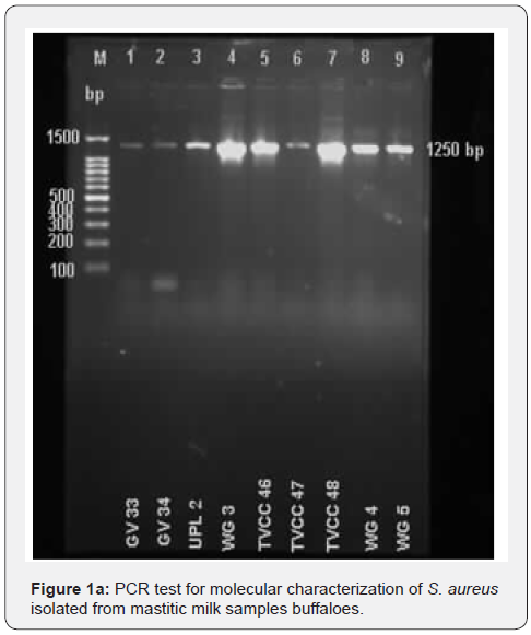
during the culture. In PCR test the S. aureus produced a specific
PCR product of 1250 bp (Figure 1a) and E. coli produced a specific
PCR product of 662 bp (Figure 1b).

In MIC assays, 31% isolates (n=13) of S. aureus were found
to be resistant to amoxicillin. The isolates were GV28, GV40,
GV42, GV43, GV45, TVCC41, TVCC47, TVCC49, TVCC53, PMNR1,
KSP35, KSP36 and KSP39. Both the plant extracts (each extract
separately) didn’t exhibit any significant antimicrobial activity
against all the isolates (n=42) of S. aureus. However, for 45.2% of
isolates (n=19) amoxicillin exhibited antimicrobial activity even at
a lower concentration when combined with the plant leaf extract.
The isolates were GV29, GV30, GV35, GV38, GV39, GV40, GV41,
GV42, GV43, GV44, GV45, TVCC42, TVCC43, TVCC47, TVCC49,
TVCC53, KSP35, KSP36 and KSP39. The MIC values of amoxicillin
in antibiotic & leaf extract combination wells are found to be
lower (to the extent of 0.00006μg/ml of concentration) than the
MIC value of amoxicillin alone. Out of 13 isolates of S. aureus that
were found to be resistant for amoxicillin, 10 isolates showed
sensitivity to amoxicillin, when it is used in combination with
the leaf extracts. For 23.8% isolates (n=10) of S. aureus there is
no significant variation in MIC values of amoxicillin, when it is
used alone or in combination with leaf extract. For 40.5% of
isolates (n=17) amoxicillin exhibited antimicrobial activity at a
lower concentration when combined with the plant root extract.
The isolates were GV30, GV40, GV41, GV42, GV43, GV44, GV45,
TVCC46, TVCC48, TVCC49, WG3, WG4, WG5, KSP35, KSP36,
KSP39 and KSP43. The MIC values of amoxicillin in antibiotic &
root extract combination wells are found to be lower than the MIC
values of amoxicillin alone. Out of 13 isolates of S. aureus that were
found to be resistant for amoxicillin, 9 isolates showed sensitivity
to amoxicillin when it is used in combination with the root extract.
In MIC assays, all the E. coli isolates (n=11) were found to be
sensitive to enrofloxacin. The isolates were GV26, GV27, GV28,
GV29, WG1, KSP35, KSP38, GV46, GV47, KSP44 and KSP45. The
leaf extract exhibited significant antimicrobial activity against
81.81% isolates (n=9) of E. coli. For these 9 isolates of E. coli
the MIC values of leaf extract were significantly lower than the
MIC values of enrofloxacin. The root aqueous extract exhibited
antimicrobial activity against all the 11 isolates of E. coli. The
MIC values of plant extracts was in the range of 31.25 to 0.98 μg/ml for different isolates of E. coli, whereas the MIC values of
enrofloxacin are in the range of 500 - 62.5 μg/ml. In MIC assays
with combination of enrofloxacin & plant extract (each extract
separately), the enrofloxacin didn’t exhibit antimicrobial activity
at its higher concentration but showed antimicrobial activity at its
lower concentration.
After 48 hr. culture the MEC attained full confluence in
tissue culture flaks and they were used for infection studies. The
cDNA obtained from different groups of MEC was quantified by
Nanodrop (Thermo) and same concentration cDNA from all the
groups was used in qRT-PCR assays.
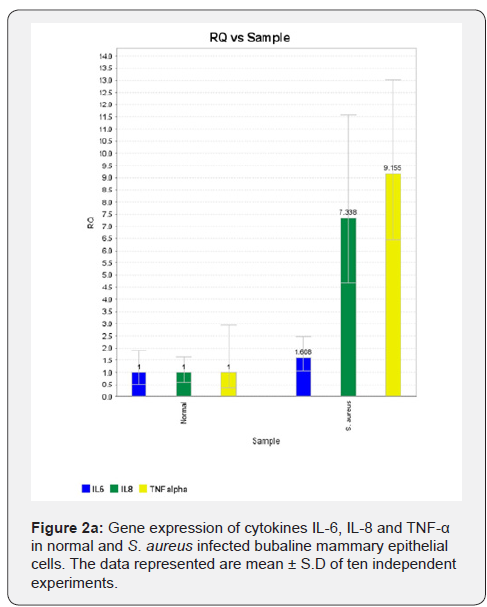
In MEC infection studies with S. aureus, the expression of
cytokines IL-6, IL-8 and TNF-α genes were upregulated in S.
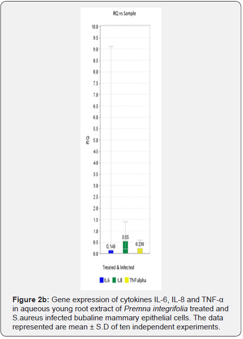
aureus infected MEC (Figure 2a). In plant (young root) extract
treated & infected MEC the gene expression of these cytokines was
significantly downregulated compared to infected MEC (Figure
2b).
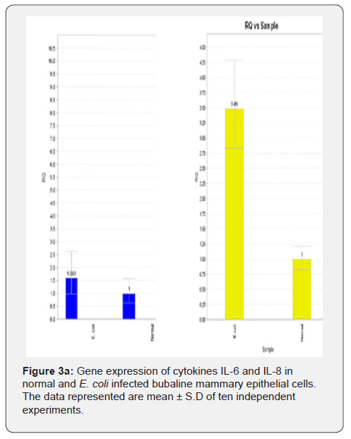
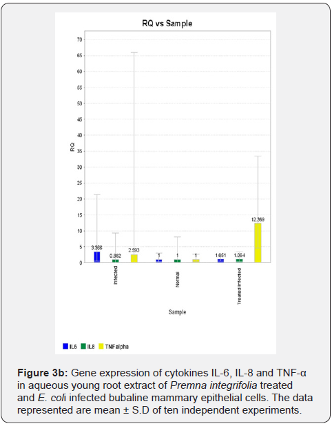
In MEC infection studies with E. coli, the gene expression
of cytokines IL-6, IL-8 and TNF-α was upregulated in infected
MEC compared to normal MEC (Figure 3a). Figure depicting
upregulation of TNF-α gene expression was not shown. The gene
expression of the cytokines IL-6 and IL-8 was downregulated
in plant (young root) extract treated & infected MEC compared
to infected MEC (Figure 3b). However, the gene expression of
cytokine TNF-α was found to be upregulated in plant (young root)
extract treated & infected MEC compared to infected MEC (Figure
3b).
Discussion
Mastitis in dairy bovines is usually caused by bacterial
pathogens leading to inflammation of udder tissue and its further
damage [1,2]. As the use of conventional antibiotics and antiinflammatory
agents have certain disadvantages like development
of antimicrobial resistance in bacteria, presence of antibiotic
residues in milk during treatment, immunosuppression associated
with cortisone administration etc., it is proposed to explore the
antibacterial and anti-inflammatory/immunomodulatory activity
of leaf and root aqueous extracts of the plant Premna integrifolia.
The antibacterial activity of the plant extracts was tested on
clinical isolates of S. aureus and E. coli isolated from mastitic
milk samples of buffaloes. The isolated S. aureus and E. coli from
different samples in this study were further characterized and the
results were in accordance with the earlier reports [2].
Out of 42 characterized isolates of S. aureus 31% showed
resistance to amoxicillin. The MIC values for indicating the
resistance to amoxicillin in S. aureus were as per the CLSI
guidelines, 2012. Due to the emergence of anti-microbial
resistance, it is not surprising to find resistance to amoxicillin
in S. aureus isolates from mastitic milk samples of dairy bovines
[4,5]. Though antibacterial activity was reported with different
extracts of Premna integrifolia [11,15-17], in the present study
both the leaf and root aqueous extracts of the plant didn’t show
any antimicrobial activity against all the isolates of S. aureus. This
may be due to use of different solvent in the process of extraction.
Also, in the previous studies the antimicrobial activity of the leaf
extract was investigated by disc diffusion method [11], whereas in
the present study the antimicrobial activity of plant extracts was
tested by micro broth dilution method. In addition, all the isolates
used in the present study were clinical isolates.
For 45.2% of isolates of S. aureus, amoxicillin exhibited
antimicrobial activity at a lower concentration when combined
with the plant leaf extract. It was reported that anti-inflammatory
drug celecoxib sensitizes S. aureus to antibiotics [12] and the
combinatorial effect of celecoxib and ampicillin was further
demonstrated [18,19]. Anti-inflammatory activity of Premna
integrifolia root was also reported [9]. Therefore, the antimicrobial
activity exhibited by amoxicillin at lower concentrations may be
due to combinatorial effect of plant extract (with anti-inflammatory
activity) and amoxicillin. This may be correlated to the finding that
out of 13 isolates of S. aureus that were found to be resistant to
amoxicillin, 10 isolates showed sensitivity to amoxicillin when
used in combination with the leaf extracts and 9 isolates showed
sensitivity to amoxicillin when used in combination with the root
extract. However, further research is to be carried out to find out
the precise mechanism of this combinatorial effect.
In MIC assays the antibiotic enrofloxacin exhibited
antimicrobial activity against all the isolates of E. coli. The
sensitivity of E. coli to enrofloxacin in antimicrobial assays was
already established [5,20]. Although the leaf extract didn’t exhibit
any significant antimicrobial activity against S. aureus isolates, it
exhibited significant antimicrobial activity against 9 isolates of
E. coli. In fact MIC values of leaf extract were significantly lower
than the MIC values of enrofloxacin for these E. coli isolates. The
antimicrobial activity exhibited by the leaf extracts against E. coli
is in accordance with the earlier reports on antibacterial activity
of Premna integrifolia [11,16,17]. The aqueous root extract of
the plant also exhibited significant antimicrobial activity against
all the 11 isolates of E. coli. Specific research reports on the
antimicrobial activity of the root extract are not available. Though
the enrofloxacin has an established antimicrobial activity against
E. coli when used alone, it is very interesting to observe in the
present study that the enrofloxacin in combination with the plant
extract (each extract separately) didn’t exhibit antimicrobial
activity at higher concentration but exhibited its antimicrobial
activity at lower concentrations. So, in two-fold serial dilution
wells of enrofloxacin with combination of constant concentration
of plant extract (each extract separately), bacterial growth was
not inhibited at higher concentrations of enrofloxacin, whereas
at lower concentrations of enrofloxacin the bacterial growth
was inhibited. However, usually in MIC assays as the dilution
of antibiotic progresses in the series of wells its concentration
decreases and the bacterial growth is not inhibited in wells of
microtiter plates with lower concentration of antibiotic. These
findings are also in contrary to the reports on synergism of natural
products and antibiotics [21].
From the studies on antimicrobial activity of
fluoroquinolone
antibiotic ciprofloxacin in combination with antioxidants it was
reported that antioxidants exhibited antagonistic activity on
ciprofloxacin [22,23]. It was observed that as the fluoroquinolones
kill the bacteria by increasing the oxidative stress in bacterial
cells, the concurrent/combinatorial use of antioxidants inhibit
the oxidative stress induced by the ciprofloxacin. The antioxidant
properties of Premna integrifolia were already reported [9-
11]. Therefore, it may be summrised that in the present study
the antioxidant properties of the plant extracts antagonized
the antimicrobial activity of enrofloxacin, which belongs to
fluoroquinolones. This is supported by the observation that with
plant extract combination E. coli growth was not inhibited in the
microtiter plate wells with higher concentration of antibiotic,
whereas in the wells with lower concentration of enrofloxacin
the E. coli growth was inhibited. Perhaps there might be optimum
levels of enrofloxacin and antioxidant plant extract combination
in the microtiter plate wells with higher concentrations of
enrofloxacin, leading to antagonistic action of plant extract on
enrofloxacin. However, further studies are required for conclusive
evidence on this aspect.
The aqueous leaf extract of the plant Premna integrifolia didn’t
have any activity on downregulation in the expression of cytokines,
IL-6, IL-8 and TNF-α genes in S. aureus and E. coli infection studies
in MEC. However, in MEC infection studies with S. aureus the
aqueous root extract of the plant Premna integrifolia showed antiinflammatory
activity by downregulating the expression of genes
of cytokines IL-6, IL-8 and TNF-α. But in MEC infection studies
with E. coli the aqueous root extract showed anti-inflammatory
activity by downregulating the expression of cytokines IL-6 and
IL-8 genes only but not TNF-α. This may be due to the potent
action of endotoxin of E. coli on MEC even after heat inactivation.
This study thus forms the first report on the pattern of expression
of cytokines IL-6, IL-8 and TNF-α genes in Premna integrifolia
plant extract treated and infected cells of any system.
Conclusion
In conclusion, although the young leaf and root extracts of the
plant Premna integrifolia didn’t exhibit any antimicrobial activity
on S. aureus, significant antimicrobial activity was exhibited by
these extracts on E. coli in microbroth dilution assays for MIC
in vitro. However, in combination with the plant extract, the
sensitivity of S. aureus to amoxicillin is not only increased but
also the S. aureus isolates that were resistant to amoxicillin also
showed sensitivity to the same antibiotic in this combination.
The effect of plant extracts on E. coli, however, were in contrast
with the findings of S. aureus as the antioxidant natural products
showed antagonism on antimicrobial activity of enrofloxacin in its
combination with the plant extracts. The aqueous root extract of
Premna integrifolia exhibited anti-inflammatory activity through
down regulation of genes of cytokines IL-6, IL-8 and TNF-α in S.
aureus infected MEC. However, the down regulation of genes of
cytokines was limited to only IL-6 and IL-8 only in E. coli infected
MEC. Therefore, the plant extracts of Premna integrifolia offer
promising solution for therapeutic approach to mastitis in dairy
animals with a caution on its antioxidant property as it antagonizes
the action of fluoroquinolone antibiotics.
Acknowledgment
The authors acknowledge the funding by National Medicinal
Plants Board (NMPB), Ministry of AYUSH, Government of India,
New Delhi to carry out this research project (Z. 18017/187/CSS/
R&D/AP-01/2014-15).


Comments
Post a Comment